Brain Aging, Brain Health, and Neurocompensatory Mechanisms in Large-Scale Brain Network Dynamics and Cognitive Function
Attention, perception, memory, and emotion
We are currently studying large-scale brain network dynamics under specific physical, anatomical constraints inferred from modern-day neuroimaging methods, such as EEG, MEG, fMRI, DTI/DWI, using resting and goal-oriented task conditions. Our group combines three complementary approaches — neuroimaging, computational methods including data-driven analysis and machine learning, and behavioural experiments grounded on neurocognitive and neuropsychological theories — to understand how multisensory perception and attentional control shape memory processing, and how cognitive aging impacts perceptual integration, attentional variability, working memory, and episodic memory processing. We also apply neurodynamical computational modelling frameworks to uncover the specific influence of biophysical and physiological parameters such as noise, conduction delay, and brain-states on the large-scale brain network structural-functional connectivity relationship (modularity, small-world network topology, scale-free topology, centrality) and dynamics (synchrony, coherence, metastability, flexibility) of the aging brain. Further, exploiting unisensory (visual, auditory, and somatosensory) and multisensory (visual-auditory, visual-tactile) fMRI and EEG paradigms, we investigate brain networks and fingerprint multisensory brain connectivity in space and time.

Majumdar et al.
Cerebral Cortex
2026 · 36(4), bhag053
Aging increases orbitofrontal neural volatility during affective inference
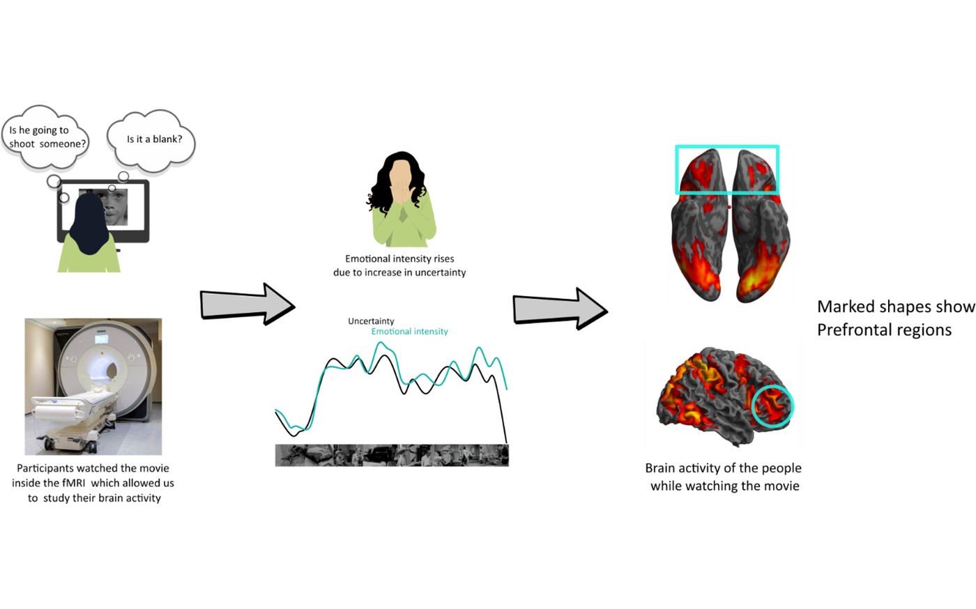
Understanding the mechanisms behind the stability and volatility of blood-oxygen-level-dependent signal is crucial in characterizing lifespan aging trajectories. Here, we propose that tracking neural fluctuations in brain areas during naturalistic tasks provides a more salient characterization of aging trajectories compared to measures based on mean and resting-state variability. Compared to other prefrontal regions, the orbitofrontal cortices exhibit higher blood-oxygen-level-dependent signal variability in aged individuals. Neural latent state analysis revealed that, in contrast to the stable representations in younger adults, the orbitofrontal cortex in older adults exhibited more temporally distorted representations, mirroring their distinct affective experiences. Furthermore, lower orbitofrontal cortex variability during the movie was associated with participants' bias toward positive responses in a separate emotional reactivity task. This suggests that orbitofrontal neural volatility might be a general adaptive response to affective inference processes. To investigate this further, a Bayesian learning model of valence dynamics was employed, which revealed that older adults exhibit heightened uncertainty in neural representations while estimating affective states. Collectively, these results indicate that neural volatility identified through blood-oxygen-level-dependent variability carries unique information about older adults' affective experiences and how naturalistic neuroimaging can chart a way forward in understanding this better.

Saha et al.
Communications Biology
2025 · 8(1), 1251
Local homeostasis preserves global neural dynamics compensating for structural loss during human lifespan aging
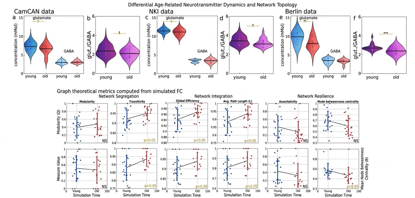
Aging brain undergoes a structural decline over lifespan accompanied by changes in neurotransmitter levels, leading to altered functional markers. Past studies have reported human resting state brain display a remarkable preservation of coordination among neural assemblies stemming from an underlying neurocomputational principles along aging trajectories, however, the true nature of which remains unknown. Here, we identify the computational mechanisms with which neurotransmitters, such as altered GABA and glutamate concentrations, can preserve functional integration across lifespan aging, despite structural decline. We employ multiscale, biophysically grounded modeling, constrained by the empirically derived anatomical connectome of the human brain, where the neurotransmitter concentrations can be free parameters that are algorithmically adjusted to maintain regional homeostasis and optimal working point. The two estimated neurotransmitters can maintain critical firing rates in the brain region and mimic age-associated functional connectivity patterns, consistent with empirical observations. We identified invariant GABA and reduced glutamate as the principle computational mechanism that can explain the topological variation of functional connectivity along lifespan, validated using graph-theoretic metrics. The results are subsequently replicated on three distinct datasets. Thus, the study offers an operational framework that integrates brain network dynamics at macroscopic and molecular scales, to gain insight into age-associated neural disorders.

Chakraborty et al.
Cerebral Cortex
2025 · 35(2)
Contributions of short- and long-range white matter tracts in dynamic compensation with aging
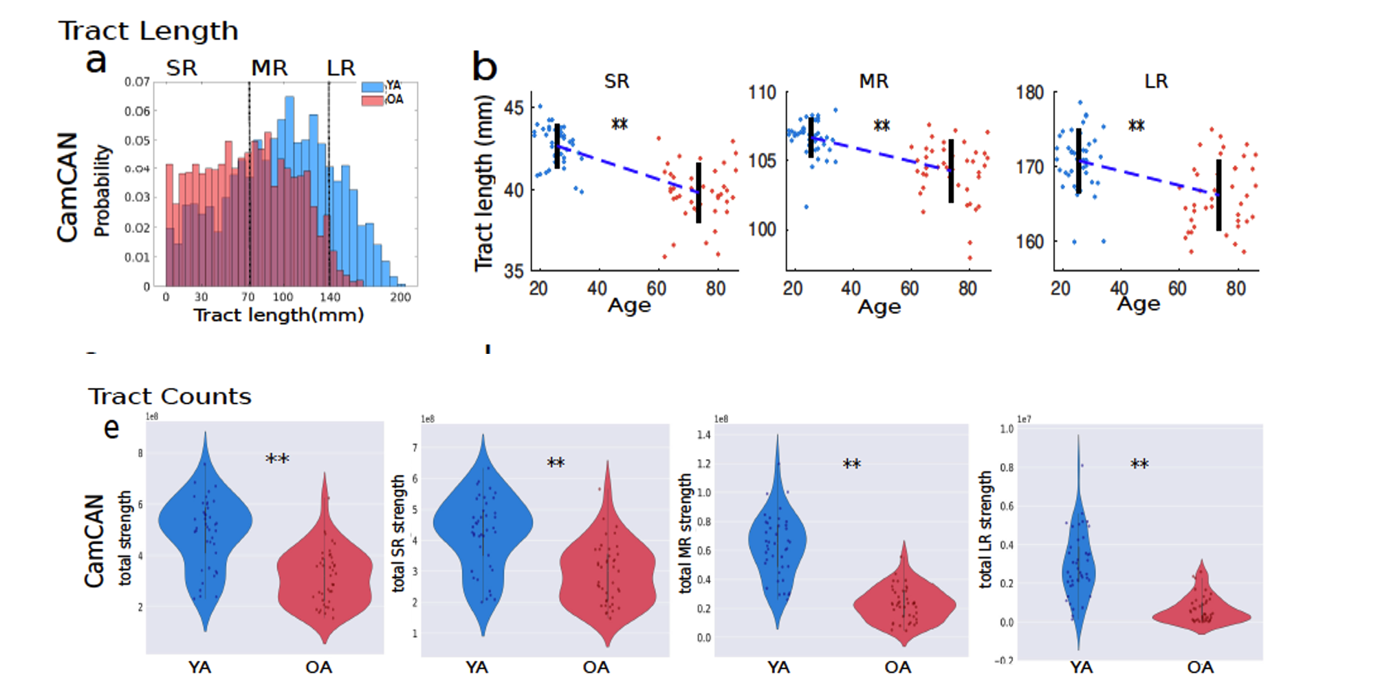
Optimal brain function is shaped by a combination of global information integration, facilitated by long-range connections, and local processing, which relies on short-range connections and underlying biological factors. With aging, anatomical connectivity undergoes significant deterioration, which affects the brain's overall function. Despite the structural loss, previous research has shown that normative patterns of functions remain intact across the lifespan, defined as the compensatory mechanism of the aging brain. However, the crucial components in guiding the compensatory preservation of the dynamical complexity and the underlying mechanisms remain uncovered. Moreover, it remains largely unknown how the brain readjusts its biological parameters to maintain optimal brain dynamics with age; in this work, we provide a parsimonious mechanism using a whole-brain generative model to uncover the role of sub-communities comprised of short-range and long-range connectivity in driving the dynamic compensation process in the aging brain. We utilize two neuroimaging datasets to demonstrate how short- and long-range white matter tracts affect compensatory mechanisms. We unveil their modulation of intrinsic global scaling parameters, such as global coupling strength and conduction delay, via a personalized large-scale brain model. Our key finding suggests that short-range tracts predominantly amplify global coupling strength with age, potentially representing an epiphenomenon of the compensatory mechanism. This mechanistically explains the significance of short-range connections in compensating for the major loss of long-range connections during aging. This insight could help identify alternative avenues to address aging-related diseases where long-range connections are significantly deteriorated.

Sastry et al.
Cerebral Cortex
2023 · 33(4), 1246-1262
Stability of sensorimotor network sculpts the dynamic repertoire of resting state over lifespan
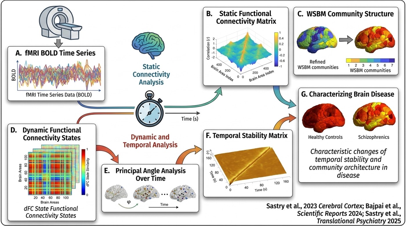
Temporally stable patterns of neural coordination among distributed brain regions are crucial for survival. Recently, many studies have highlighted the association between healthy aging and modifications in the organization of functional brain networks across various time-scales. Nonetheless, the quantitative characterization of the temporal stability of functional brain networks across healthy aging remains unexplored. This study introduces a data-driven unsupervised approach to capture high-dimensional dynamic functional connectivity (dFC) via low-dimensional patterns and subsequent estimation of temporal stability using quantitative metrics. Healthy aging-related changes in temporal stability of dFC were characterized across resting-state, movie-viewing, and sensorimotor tasks (SMT) on a large (n = 645) healthy aging dataset (18–88 years). Prominent results reveal that (1) whole-brain temporal dynamics of the dFC movie-watching task are closer to resting-state than to SMT, with an overall trend of highest temporal stability observed during SMT, followed by movie-watching and resting-state, invariant across lifespan aging, (2) in both task conditions, stability of neurocognitive networks in young adults is higher than older adults, and (3) temporal stability of whole-brain resting-state follows a U-shaped curve along lifespan—a pattern shared by sensorimotor network stability, indicating their deeper relationship. Overall, the results can be applied generally for studying cohorts of neurological disorders using neuroimaging tools.

Pathak et al.
Communications Biology
2022 · 5(1), 567
Biophysical mechanism underlying compensatory preservation of neural synchrony over the adult lifespan
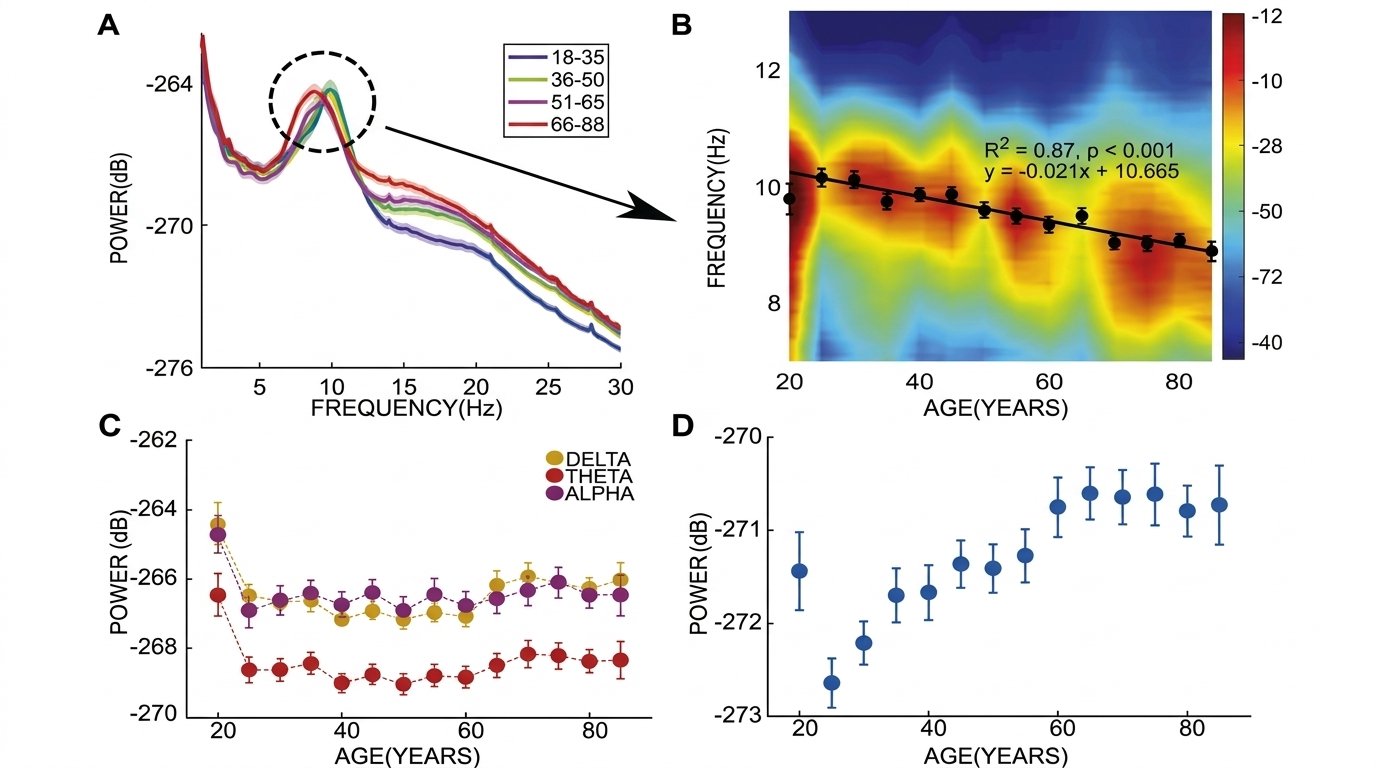
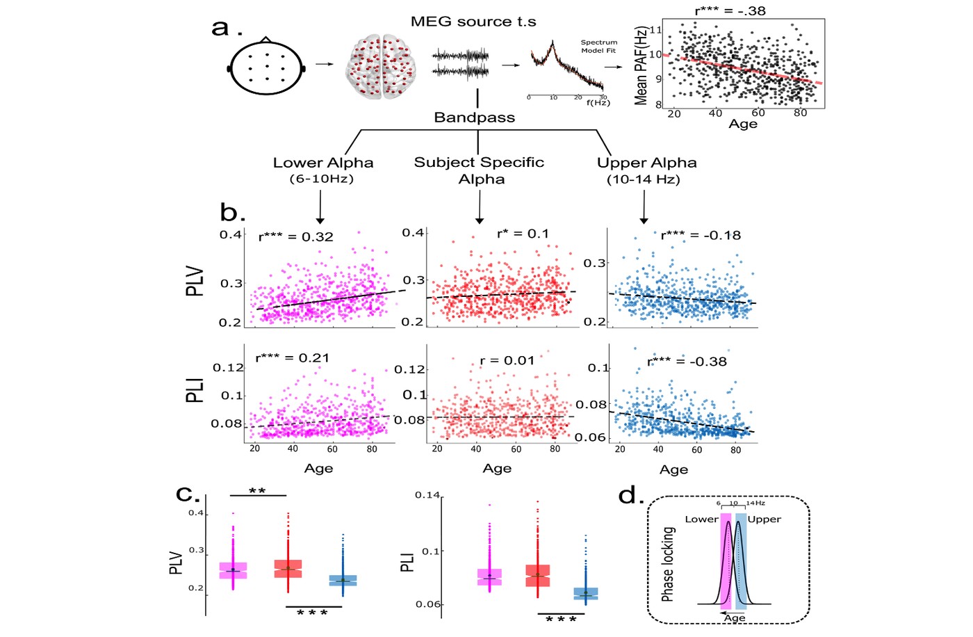
This work proposes that the preservation of functional integration, estimated from measures of neural synchrony, is a key objective of neurocompensatory mechanisms associated with healthy human ageing. To support this proposal, we demonstrate how phase-locking at the peak alpha frequency in Magnetoencephalography recordings remains invariant over the lifespan in a large cohort of human participants, aged 18-88 years. Using empirically derived connection topologies from diffusion tensor imaging data, we create an in-silico model of whole-brain alpha dynamics. We show that enhancing inter-areal coupling can cancel the effect of increased axonal transmission delays associated with age-related degeneration of white matter tracts, albeit at slower network frequencies. By deriving analytical solutions for simplified connection topologies, we further establish the theoretical principles underlying compensatory network re-organization. Our findings suggest that frequency slowing with age- frequently observed in the alpha band in diverse populations- may be viewed as an epiphenomenon of the underlying compensatory mechanism.

Das et al.
Cerebral Cortex
2021 · 31(4), 1970-1862
Reconfiguration of directed functional connectivity among neurocognitive networks with aging: considering the role of thalamo-cortical interactions
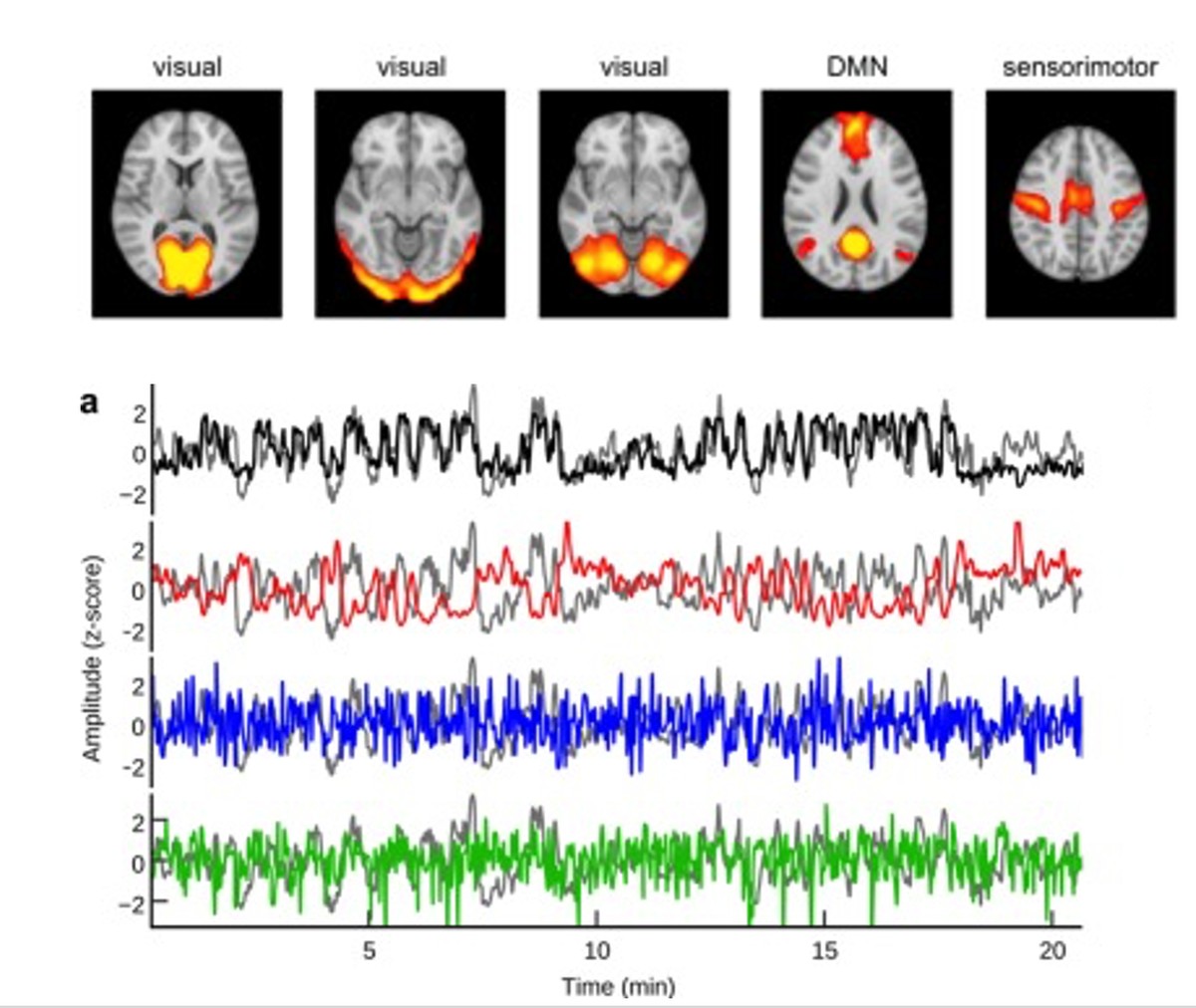
A complete picture of how subcortical nodes, such as the thalamus, exert directional influence on large-scale brain network interactions across age remains elusive. Using directed functional connectivity and weighted net causal outflow on resting-state fMRI data, we provide evidence of a comprehensive reorganization within and between neurocognitive networks (default mode: DMN, salience: SN, and central executive: CEN) associated with age and thalamocortical interactions. We hypothesize that the thalamus subserves both modality-specific and integrative hub roles in organizing causal weighted outflow among large-scale neurocognitive networks. To this end, we observe that within-network directed functional connectivity is driven by the thalamus and progressively weakens with age. Secondly, we find that the age-associated increase in between CEN- and DMN-directed functional connectivity is driven by both the SN and the thalamus. Furthermore, left and right thalami act as a causal integrative hub exhibiting substantial interactions with neurocognitive networks with aging and play a crucial role in reconfiguring network outflow. Notably, these results were largely replicated on an independent dataset of matched young and old individuals. Our findings strengthen the hypothesis that the thalamus is a key causal hub balancing both within- and between-network connectivity associated with age and maintenance of cognitive functioning with aging.

Thuwal et al.
eNeuro
2021 · 8(5)
Aperiodic and periodic components of ongoing oscillatory brain dynamics link distinct functional aspects of cognition across the adult lifespan
Signal transmission in the brain propagates via distinct oscillatory frequency bands, but the aperiodic component, 1/f activity, almost always co-exists, which most of the previous studies have not sufficiently taken into consideration. We used a recently proposed parameterization model that delimits the oscillatory and aperiodic components of neural dynamics on lifespan aging data collected from human participants using magnetoencephalography (MEG). Since healthy aging underlines an enormous change in local tissue properties, any systematic relationship of 1/f activity would highlight their impact on the self-organized critical functional states. Furthermore, we have used patterns of correlation between aperiodic background and metrics of behavior to understand the domain-general effects of 1/f activity. We suggest that age-associated global change in 1/f baseline alters the functional critical states of the brain, affecting the global information processing, impacting all aspects of cognition critically, e.g., metacognitive awareness, speed of retrieval of memory, cognitive load, and accuracy of recall through the adult lifespan. This alteration in 1/f crucially impacts the oscillatory features' peak frequency (PF) and band power ratio, which relates to more local processing and selective functional aspects of cognitive processing during the visual short-term memory (VSTM) task. In summary, this study, leveraging on big lifespan data for the first time, tracks the cross-sectional lifespan-associated periodic and aperiodic dynamical changes in the resting state to demonstrate how normative patterns of 1/f activity, PF, and band ratio (BR) measures provide distinct functional insights about the cognitive decline through adult lifespan.

Sahoo et al.
NeuroImage
2020 · 216, 116824
Lifespan-associated changes in global patterns of coherent communication
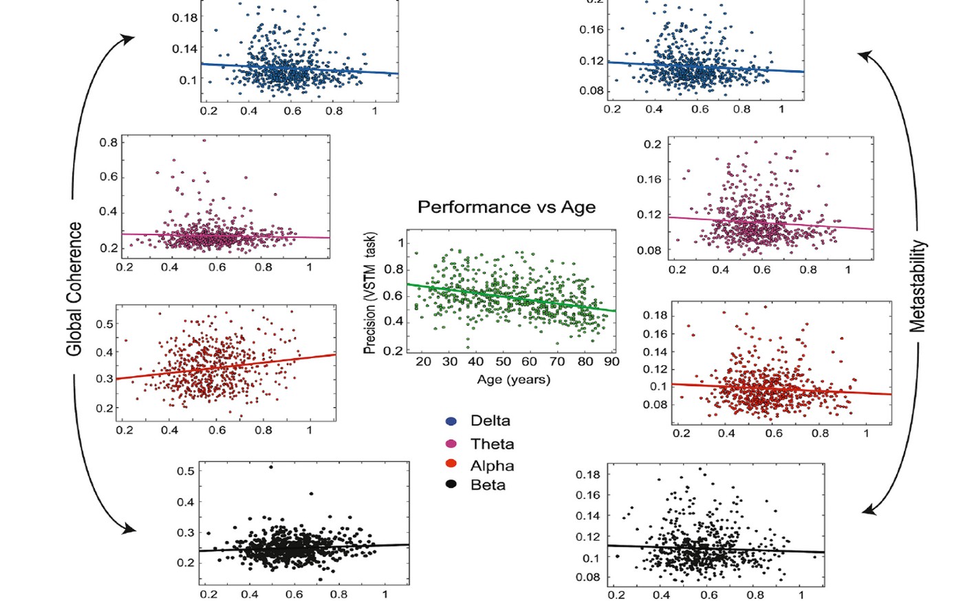
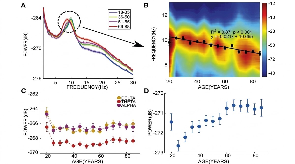
Healthy ageing is accompanied by changes to spontaneous electromagnetic oscillations. At the macroscopic scale, previous studies have quantified the basic features, e.g., power and frequencies in rhythms of interest from the perspective of attention, perception, learning, and memory. On the other hand, signatures and modes of neural communication have recently been argued to be identifiable from global measures applied on neuro-electromagnetic data, such as global coherence that quantifies the degree of togetherness of distributed neural oscillations and metastability that parametrizes the transient dynamics of the network switching between successive stable states. Here, we demonstrate that global coherence and metastability can be informative measures to track healthy ageing dynamics over lifespan, and together with the traditional spectral measures provide an attractive explanation of neuronal information processing. Finding normative patterns of brain rhythms in resting state MEG would naturally pave the way for tracking task-relevant metrics that could crucially determine cognitive flexibility and performance. While previously reported observations of a reduction in peak alpha frequency and increased beta power in older adults are reflective of changes at individual sensors (during rest and task), global coherence and metastability pinpoint the underlying coordination dynamics over multiple brain areas across the entire lifespan. In addition to replication of the previous observations in a substantially larger lifespan cohort than what was previously reported, we also demonstrate, for the first time to the best of our knowledge, age-related changes in coherence and metastability in signals over time scales of neuronal processing. Furthermore, we observed a marked frequency dependence in changes in global coordination dynamics, which, coupled with the long-held view of specific frequency bands subserving different aspects of cognition, hints at differential functional processing roles for slower and faster brain dynamics.

Naik et al.
Trends in Cognitive Sciences
2017
Metastability in senescence
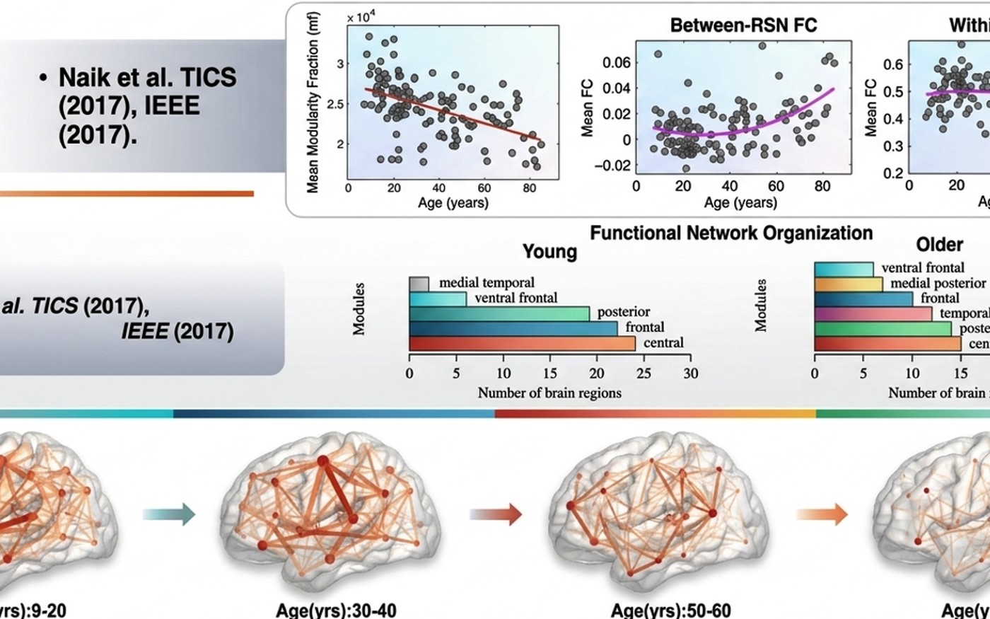
Grow old along with me! The best is yet to be, the last of life, for which the first was made (Robert Browning). Senescence has always been associated with inevitable physical and psychological deterioration. However, years of scientific scrutiny of this idea have revealed that age-related changes are not straightforward. While some of the skills are compromised with age, others remain intact or even improve with age [1]. One of the challenges of the studies on aging is therefore to solve the prediction and changes of Metastability in the Aging Brain. The notion of metastability can be implemented at two distinct timescales, one at the level of the lifespan and the other at the scale of an empirical observation, for example, a brain-scanning session of a few minutes during rest. Undoubtedly, changes in the brain that unfold at the scale of empirical observation will have repercussions for lifespan data, and this has been the argument for several of the studies that compartmentalize aging-related structural and functional changes. However, the existing Theories of Cognitive Aging: Compensation or Dedifferentiation remains an open question. The normal process of aging results in volumetric changes in grey and white matter, topological changes in the white matter fiber network, and organizational changes in functional brain networks (Box 1). For brevity, we ignore the genetic and molecular changes that might affect the observed changes [35]. Age effects across the brain regions and across cognitive modalities are not uniform, and regions such as the prefrontal cortex, the medial temporal cortex, and other heteromodal associative regions. Here, we provide an avenue to track age-associated alterations in metrics of Metastability to the Mechanisms of Aging. A growing body of research points to age-related changes in brain signal variability 17, 18, 49. The message from these studies seems to be that greater signal variability is exhibited by high-performing young adults compared to older adults across different types of tasks. Further, the correlation between BOLD signal variability and cognitive performance differs across brain regions. Metrics that quantify the variability at the network level (such as FCD) would successfully capture the aging brain dynamics and their stability over time. All thoughts and cognitive operations emerge from the stream of consciousness − a widely held belief of the Western and Eastern philosophies. In the context of the observable brain dynamics, there is no permanent state of the brain that sustains across the lifespan, and it is the transient brain dynamics that best capture the aging brain dynamics.
Mapping animal-human behavioural responses under social cues and naturalistic experience by decoding neural activity
Atypical neurodevelopment in children — fMRI, EEG, and naturalistic tasks

Bhavna et al.
Frontiers in Neuroinformatics
2024 · 18, 1392661
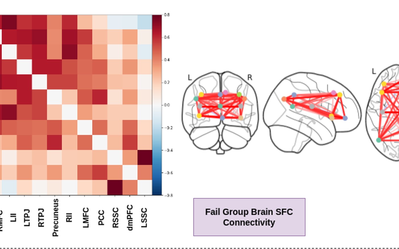
Explainable deep-learning framework: decoding brain states and prediction of individual performance in false-belief task at early childhood stage
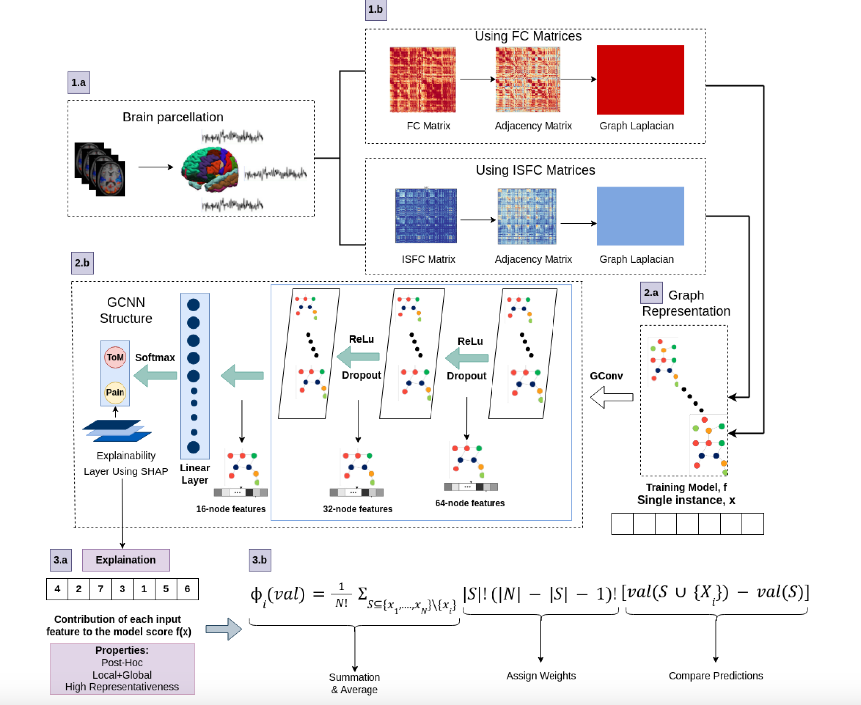
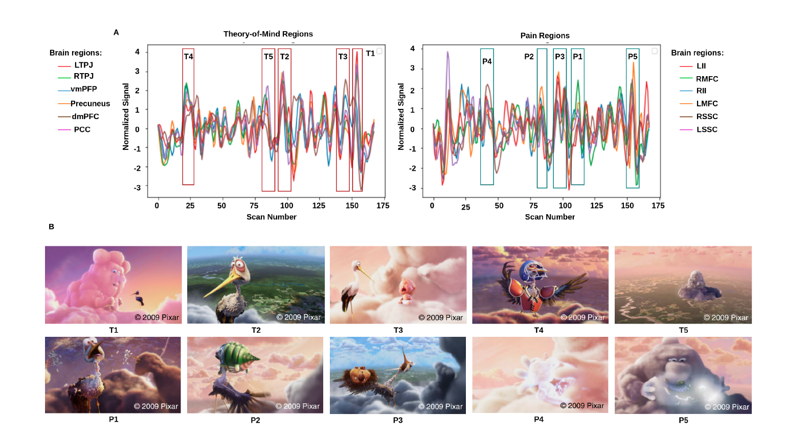
Decoding of cognitive states aims to identify individuals' brain states and brain fingerprints to predict behavior. Deep learning provides an important platform for analyzing brain signals at different developmental stages to understand brain dynamics. Due to their internal architecture and feature extraction techniques, existing machine-learning and deep-learning approaches are suffering from low classification performance and explainability issues that must be improved. In the current study, we hypothesized that even at the early childhood stage (as early as 3-years), connectivity between brain regions could decode brain states and predict behavioral performance in false-belief tasks. To this end, we proposed an explainable deep learning framework to decode brain states (Theory of Mind and Pain states) and predict individual performance on ToM-related false-belief tasks in a developmental dataset. We proposed an explainable spatiotemporal connectivity-based Graph Convolutional Neural Network (Ex-stGCNN) model for decoding brain states. Here, we consider a developmental dataset, N = 155 (122 children; 3–12 yrs and 33 adults; 18–39 yrs), in which participants watched a short, soundless animated movie, shown to activate Theory-of-Mind (ToM) and pain networks. After scanning, the participants underwent a ToM-related false-belief task, leading to categorization into the pass, fail, and inconsistent groups based on performance. We trained our proposed model using Functional Connectivity (FC) and Inter-Subject Functional Correlations (ISFC) matrices separately. We observed that the stimulus-driven feature set (ISFC) could capture ToM and Pain brain states more accurately, with an average accuracy of 94%, whereas it achieved 85% accuracy using FC matrices. We also validated our results using five-fold cross-validation and achieved an average accuracy of 92%. Besides this study, we applied the SHapley Additive exPlanations (SHAP) approach to identify brain fingerprints that contributed the most to predictions. We hypothesized that ToM network brain connectivity could predict individual performance on false-belief tasks. We proposed an Explainable Convolutional Variational Auto-Encoder (Ex-Convolutional VAE) model to predict individual performance on false-belief tasks and trained the model using FC and ISFC matrices separately. ISFC matrices again outperformed the FC matrices in the prediction of individual performance. We achieved 93.5% accuracy with an F1-score of 0.94 using ISFC matrices and achieved 90% accuracy with an F1-score of 0.91 using FC matrices.
Bhavna et al.
Scientific Reports
2024 · 14(1), 22479
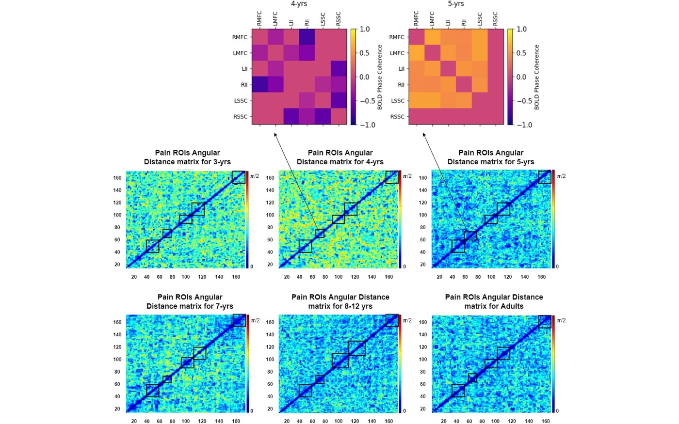
Characterization of the temporal stability of ToM and pain functional brain networks carrying distinct developmental signatures during naturalistic viewing
A temporally stable functional brain network pattern among coordinated brain regions is fundamental to stimulus selectivity and functional specificity during the critical period of brain development. Brain networks that are recruited in time to process internal states of others' bodies (like hunger and pain) versus internal mental states (like beliefs, desires, and emotions) of others' minds allow us to ask whether a quantitative characterization of the stability of these networks carries meaning during early development and constrain cognition in a specific way. Previous research provides critical insight into the early development of the theory-of-mind (ToM) network and its segregation from the Pain network throughout normal development using functional connectivity. However, a quantitative characterization of the temporal stability of ToM networks from early childhood to adulthood remains unexplored. In this work, reusing a large sample of children (n = 122, 3–12 years) and adults (n = 33) dataset that is available on the OpenfMRI database under the accession number ds000228, we addressed this question based on their fMRI data during a short and engaging naturalistic movie-watching task. The movie highlights the characters' bodily sensations (often pain) and mental states (beliefs, desires, emotions), and is a feasible experiment for young children. Our results tracked the change in temporal stability using an unsupervised characterization of ToM and Pain networks DFC patterns using Angular and Mahalanobis distances between dominant dynamic functional connectivity subspaces. Our findings reveal that both ToM and Pain networks exhibit lower temporal stability as early as 3-years and gradually stabilize by 5-years, which continues till adolescence and late adulthood (often sharing similarity with adult DFC stability patterns). Furthermore, we find that the temporal stability of ToM brain networks is associated with the performance of participants in the false belief task to access mentalization at an early age. Interestingly, higher temporal stability is associated with the pass group, and similarly, moderate and low temporal stability are associated with the inconsistent group and the fail group. Our findings open an avenue for applying the temporal stability of large-scale functional brain networks during cortical development to act as a biomarker for multiple developmental disorders concerning impairment and discontinuity in the neural basis of social cognition.

Nair et al.
2022 · 16, 878046
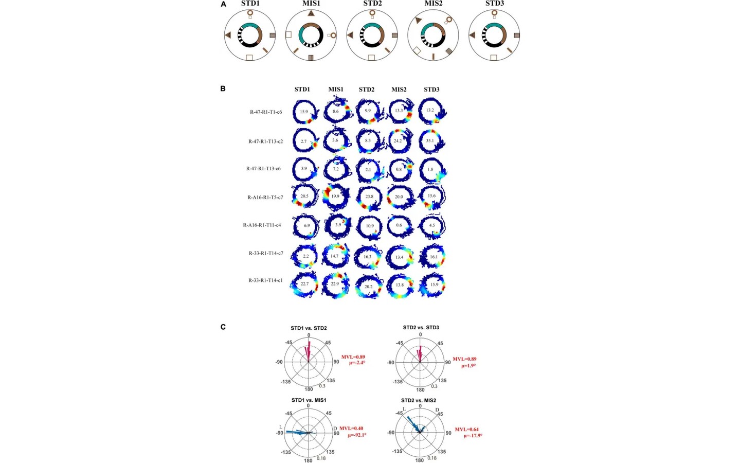
Hippocampus maintains a coherent map under reward feature-landmark cue conflict
Animals predominantly use salient visual cues (landmarks) for efficient navigation. When the relative position of the visual cues is altered, the hippocampal population exhibits heterogeneous responses and constructs context-specific spatial maps. Another critical factor that can strongly modulate spatial representation is the presence of reward. Reward features can drive behavior and are known to bias spatial attention. However, it is unclear whether reward features are used for spatial reference in the presence of distal cues and how the hippocampus population dynamics change when the association between reward features and distal cues is altered. We systematically investigated these questions by recording place cells from the CA1 in different sets of experiments while the rats ran in an environment with the conflicting association between reward features and distal cues. We report that, when reward features were only used as local cues, the hippocampal place fields exhibited coherent and dynamical orientation across sessions, suggesting the use of a single coherent spatial map. We found that place cells maintained their spatial offset in the cue conflict conditions, thus showing a robust spatial coupling featuring an attractor-like property in the CA1. These results indicate that reward features may control the place field orientation but may not cause sufficient input difference to create context-specific spatial maps in the CA1.
Atypical Brain Network Dynamics in Neurodevelopmental Disorders
Neurophysiological processes and behavioral responses in human subjects are measurable indirectly using fMRI, and directly using surface recordings such as EEG and MEG — brain oscillations, normal responses that change with cortical lesion, learning, and memory consolidation, and that interact dynamically with the intrinsic spontaneous oscillations present in the brain due to large-scale anatomy and connectivity between brain modules in the absence of external stimuli. We are interested in specific alterations in cognitive response and the neuronal changes that underlie them. To address this, we systematically develop mathematical tools for understanding how brain networks reconfigure over multiple time scales, and apply these tools with our collaborators to understand perceptual learning, vision, and psychiatric disease.
Bhavna et al.
Network Neuroscience
2025 · 1-29
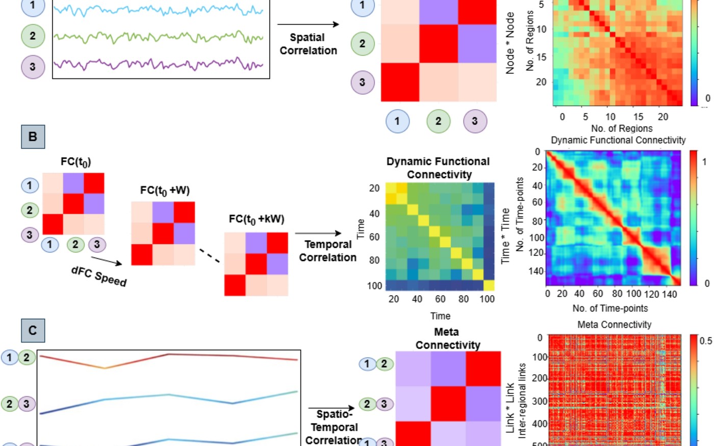
A lightweight, end-to-end explainable, and generalized attention-based graph neural network model trained on high-order spatiotemporal organization of dynamic functional connectivity to classify autistics from typically developing
Autism spectrum disorder (ASD) is a neurodevelopmental disorder characterized by deficits in social cognition, interaction, communication, restricted behaviors, and sensory abnormalities. The heterogeneity in ASD's clinical presentation complicates its diagnosis and treatment. Recent technological advancements in graph neural networks (GNNs) have been extensively used to diagnose brain disorders such as ASD, but existing machine learning models often suffer from low accuracy and explainability. In this study, we proposed a novel, explainable, and generalized node-edge connectivity-based graph attention neural network (Ex-NEGAT) model, leveraging edge-centric high-order spatiotemporal organization of dynamic functional connectivity streams between large-scale functional brain networks implicated in autism. Using the Autism Brain Imaging Data Exchange I and II datasets (total samples = 1,500), the model achieved 88% accuracy and an F1-score of 0.89. Additionally, we used meta-connectivity subtypes to identify subgroups within ASD samples using the rough fuzzy c-means algorithm. We also used connectome-based prediction modeling, which revealed critical brain networks contributing to predictions that accurately correlate with Autism Diagnostic Observation Schedule (ADOS) and full intelligence quotient (FIQ) scores. The proposed framework offers a robust approach based on previously unexplored higher-order spatiotemporal correlation features of dynamic functional connectivity, which may provide critical insight into ASD heterogeneity and improve diagnostic precision.

Sigar et al.
Autism Research
2023 · 16(1), 66-83
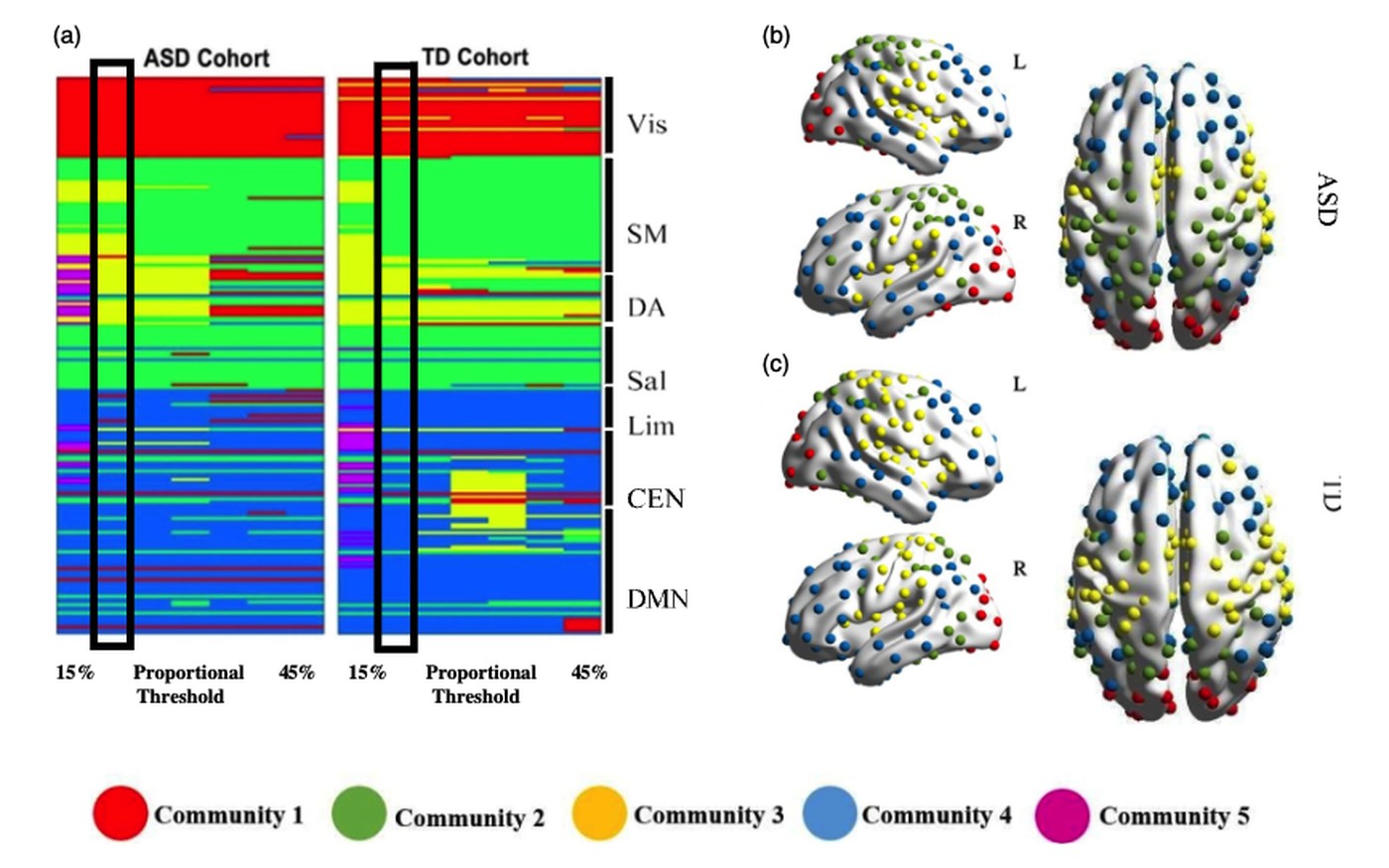
Altered global modular organization of intrinsic functional connectivity in autism arises from atypical node-level processing
Autism spectrum disorder (ASD) is a neurodevelopmental condition characterized by restricted interests and repetitive behaviors as well as social-communication deficits. These traits are associated with atypicality of functional brain networks. Modular organization in the brain plays a crucial role in network stability and adaptability for neurodevelopment. Previous neuroimaging research demonstrates discrepancies in studies of functional brain modular organization in ASD. These discrepancies result from the examination of mixed age groups. Furthermore, recent findings suggest that while much attention has been given to deriving atlases and measuring the connections between nodes, within-node information may also be crucial in determining altered modular organization in ASD compared with typical development (TD). However, altered modular organization originating from systematic nodal changes is yet to be explored in younger children with ASD. Here, we used graph-theoretical measures to fill this knowledge gap. To this end, we utilized multicenter resting-state fMRI data collected from 5 to 10-year-old children, 34 with ASD and 40 TD, obtained from the Autism Brain Image Data Exchange (ABIDE) I and II. We demonstrate that alterations in topological roles and modular cohesiveness are the two key properties of brain regions anchored in default mode, sensorimotor, and salience networks, and primarily relate to social and sensory deficits in children with ASD. These results demonstrate that atypical global network organization in children with ASD arises from nodal role changes, and contribute to the growing body of literature suggesting that there is interesting information within nodes providing critical markers of functional brain networks in autistic children.

Roy et al.
Network Neuroscience
2021 · 1-27
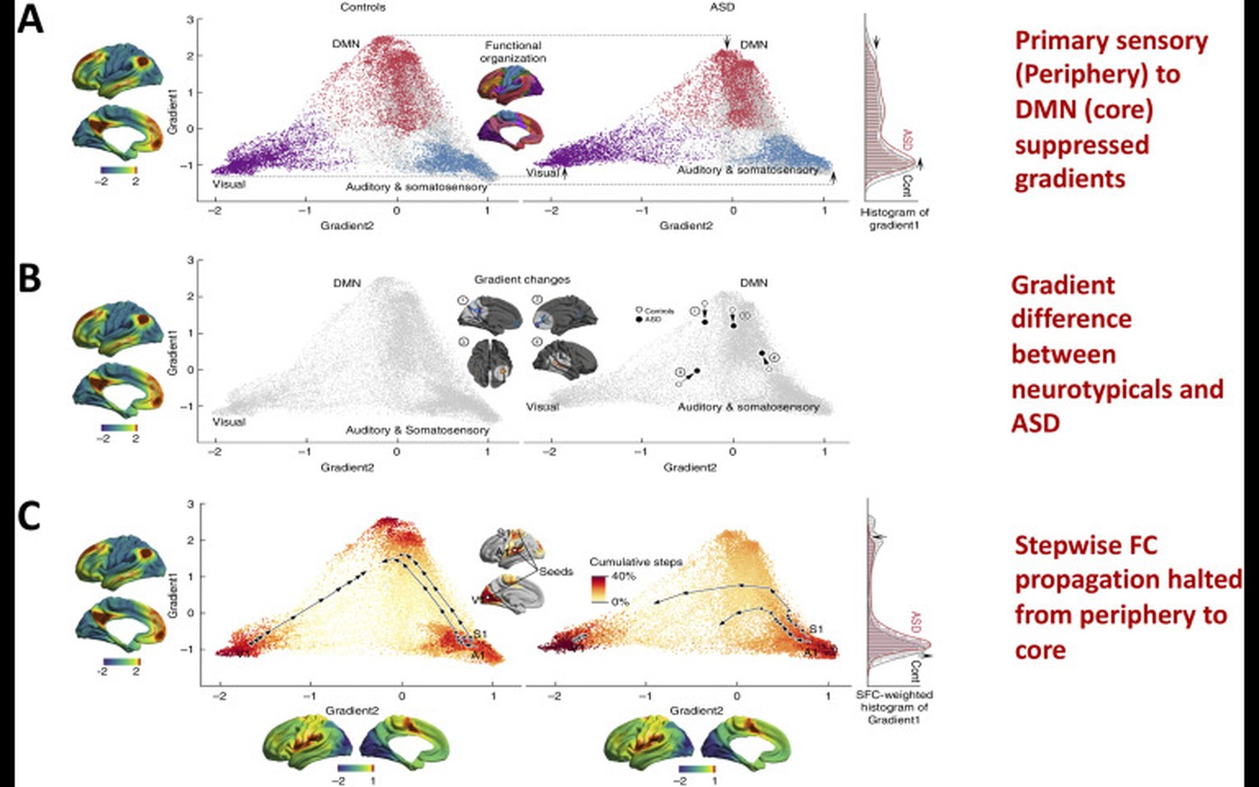
Atypical core-periphery brain dynamics in autism: implications for symptom severity
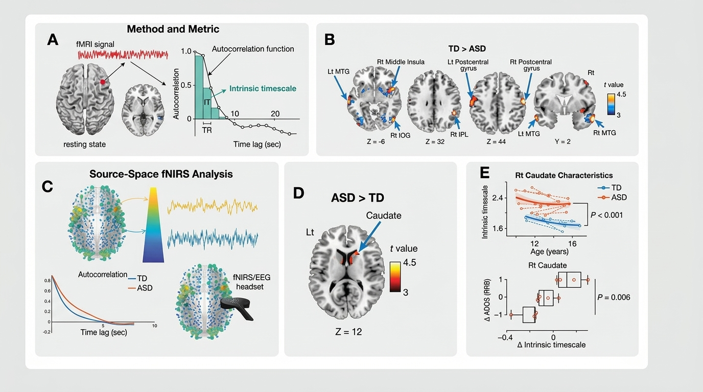
The intrinsic function of the human brain is dynamic, giving rise to numerous behavioral subtypes that fluctuate distinctively at multiple timescales. One of the key dynamical processes that takes place in the brain is the interaction between core-periphery brain regions, which undergoes constant fluctuations associated with developmental time frames. Core-periphery dynamical changes associated with macroscale brain network dynamics span multiple timescales and may lead to atypical behavior and clinical symptoms. For example, recent evidence suggests that brain regions with shorter intrinsic timescales are located at the periphery of brain networks (e.g., sensorimotor hand, face areas) and are implicated in perception and movement. On the contrary, brain regions with longer timescales are core hub regions. These hubs are important for regulating interactions between the brain and the body during self-related cognition and emotion. In this review, we summarize a large body of converging evidence derived from time-resolved fMRI studies in autism to characterize atypical core-periphery brain dynamics and how they relate to core and contextual sensory and cognitive profiles.
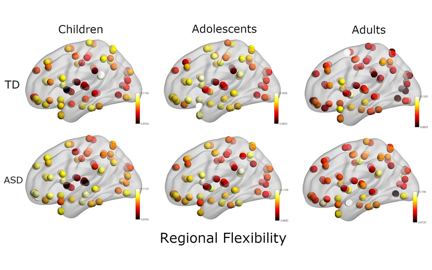
Atypical flexibility in dynamic functional connectivity quantifies the severity in autism spectrum disorder
Resting-state functional connectivity (FC) analyses have shown atypical connectivity in autism spectrum disorder (ASD) as compared to typically developing (TD). However, this view emerges from investigating static FC overlooking the whole brain transient connectivity patterns. In our study, we investigated how age and disease influence the dynamic changes in functional connectivity of TD and ASD. We used resting-state functional magnetic resonance imaging (rs-fMRI) data stratified into three cohorts: children (7–11 years), adolescents (12–17 years), and adults (18+ years) for the analysis. The dynamic variability in the connection strength and the modular organization in terms of measures such as flexibility, cohesion strength, and disjointness were explored for each subject to characterize the differences between ASD and TD. In ASD, we observed significantly higher inter-subject dynamic variability in connection strength as compared to TD. This hyper-variability relates to the symptom severity in ASD. We also found that whole-brain flexibility correlates with static modularity only in TD. Further, we observed a core-periphery organization in the resting-state, with Sensorimotor and Visual regions in the rigid core; and DMN and attention areas in the flexible periphery. TD also develops a more cohesive organization of sensorimotor areas. However, in ASD, we found a strong positive correlation of symptom severity with flexibility of rigid areas and with disjointness of sensorimotor areas. The regions of the brain showing high predictive power of symptom severity were distributed across the cortex, with stronger bearings in the frontal, motor, and occipital cortices. Our study demonstrates that the dynamic framework best characterizes the variability in ASD.
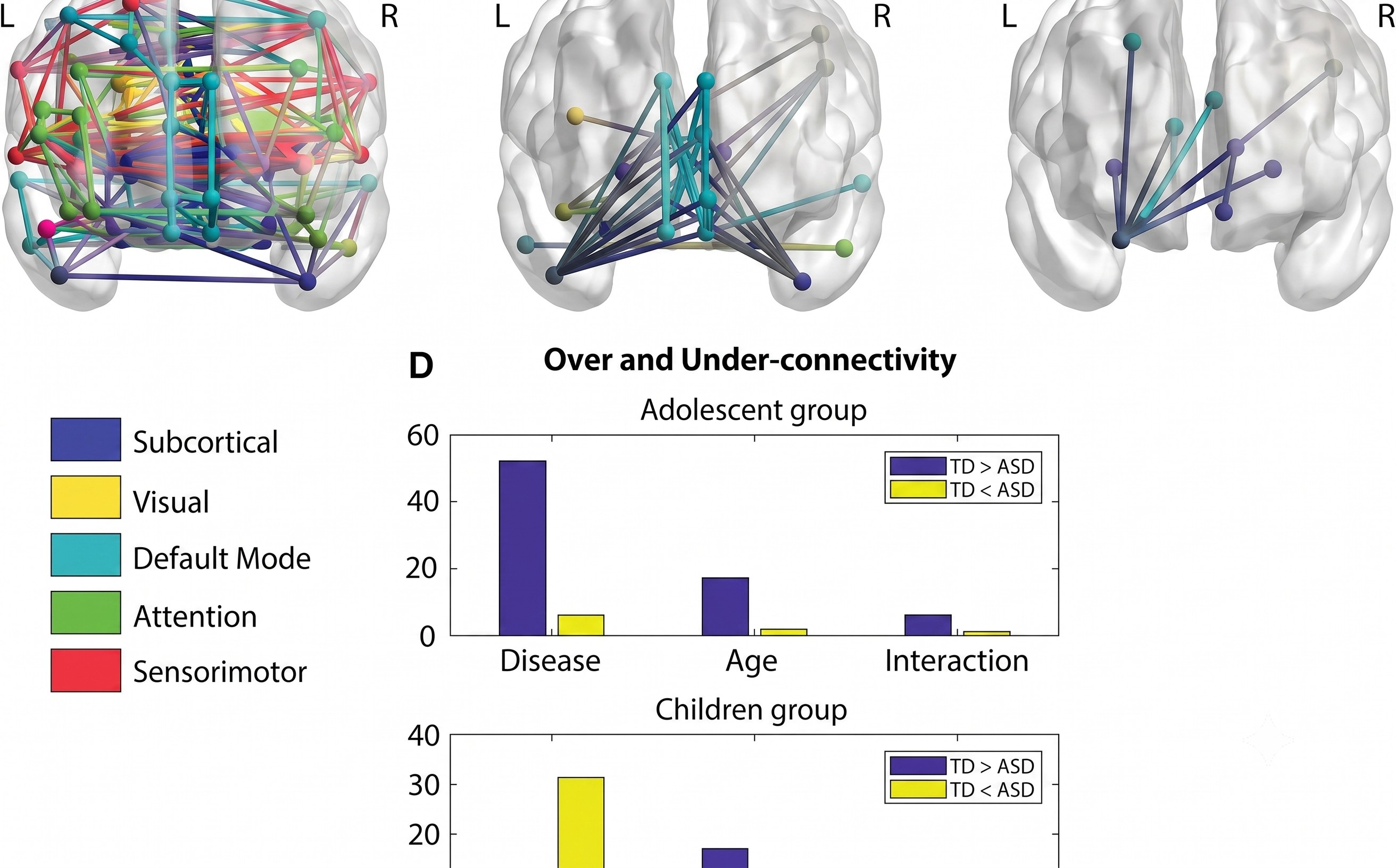

Harlalka et al.
Brain Connectivity
2018 · 8(7)
Age, disease, and their interaction effects on the intrinsic connectivity of children and adolescents in Autism Spectrum Disorder using functional connectomics
Brain connectivity analysis has provided crucial insights to pinpoint the differences between autistic and typically developing (TD) children during development. This study aims to investigate the functional connectomics of autism spectrum disorder (ASD) versus TD and to underpin the effects of development, disease, and their interactions on the observed atypical brain connectivity patterns. Resting-state functional magnetic resonance imaging (rs-fMRI) from the Autism Brain Imaging Data Exchange (ABIDE) data set, which is stratified into two cohorts: children (9-12 years) and adolescents (13-16 years), is used for the analysis. Differences in various graph theoretical network measures are calculated between ASD and TD in each group. Furthermore, a two-factor analysis of variance test is used to study the effect of age, disease, and their interaction on the network measures and the network edges. Furthermore, the differences in connection strength between TD and ASD subjects are assessed using network-based statistics. The results showed that ASD exhibits increased functional integration at the expense of decreased functional segregation. In ASD adolescents, there is a significant decrease in modularity, suggesting a less robust modular organization, and an increase in participation coefficient, suggesting more random integration and widely distributed connection edges. Furthermore, there is significant hypoconnectivity observed in the adolescent group, especially in the default mode network, while the children group shows both hyper- and hypoconnectivity. This study lends support to a model of global atypical connections and further identifies functional networks and areas that are independently affected by age, disease, and their interaction.
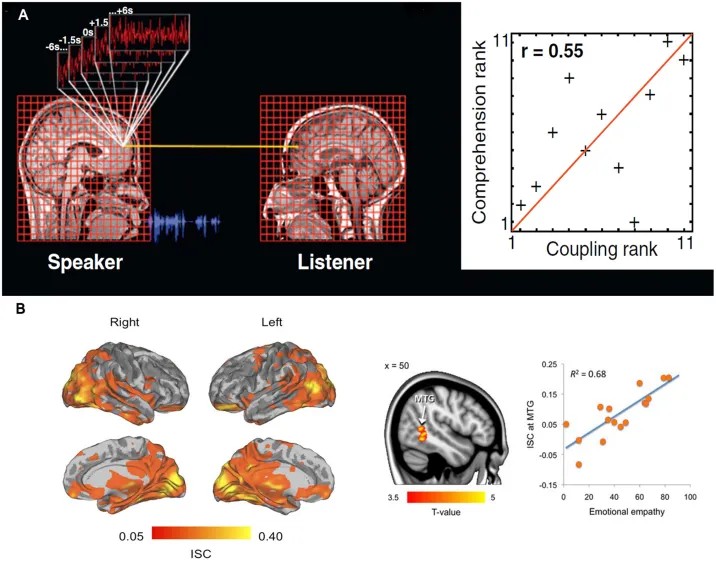

Ray et al.
Frontiers in Psychology
2017
The neural substrate of group mental health: insights from a multi-brain reference frame in functional neuroimaging
Contemporary mental health practice primarily centers around the neurobiological and psychological processes at the individual level. However, a more careful consideration of interpersonal and other group-level attributes (e.g., interpersonal relationships, mutual trust/hostility, interdependence, and cooperation) and a better grasp of their pathology can add a crucial dimension to our understanding of mental health problems. A few recent studies have explored the interpersonal behavioral processes in the context of various psychiatric disorders. Neuroimaging can supplement these approaches by providing insight into the neurobiology of interpersonal functioning. Keeping this view in mind, we discuss a recently developed approach in functional neuroimaging that calls for a shift from a focus on neural information contained within brain space to a multi-brain framework exploring the degree of similarity/dissimilarity of neural signals between multiple interacting brains. We hypothesize novel applications of quantitative neuroimaging markers, such as inter-subject correlation, that may be able to evaluate the role of interpersonal attributes affecting an individual or a group. Empirical evidence of the usage of these markers in understanding the neurobiology of social interactions is provided to argue for their application in future mental health research.
Multi-Scale, Multimodal Imaging and Machine Learning to Characterize Brain Lesions and Axonal Injury
Structure-function-dynamics in cognitive processing and functional recovery
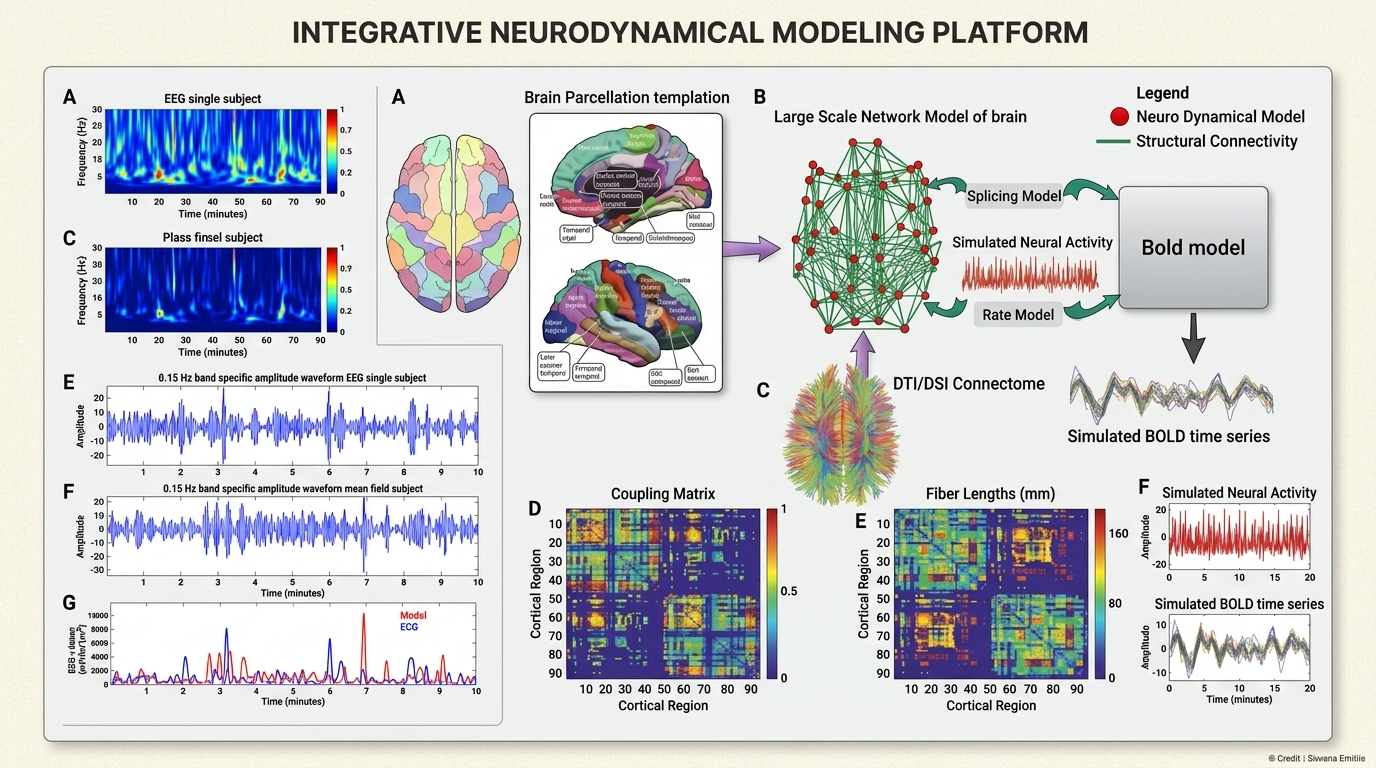
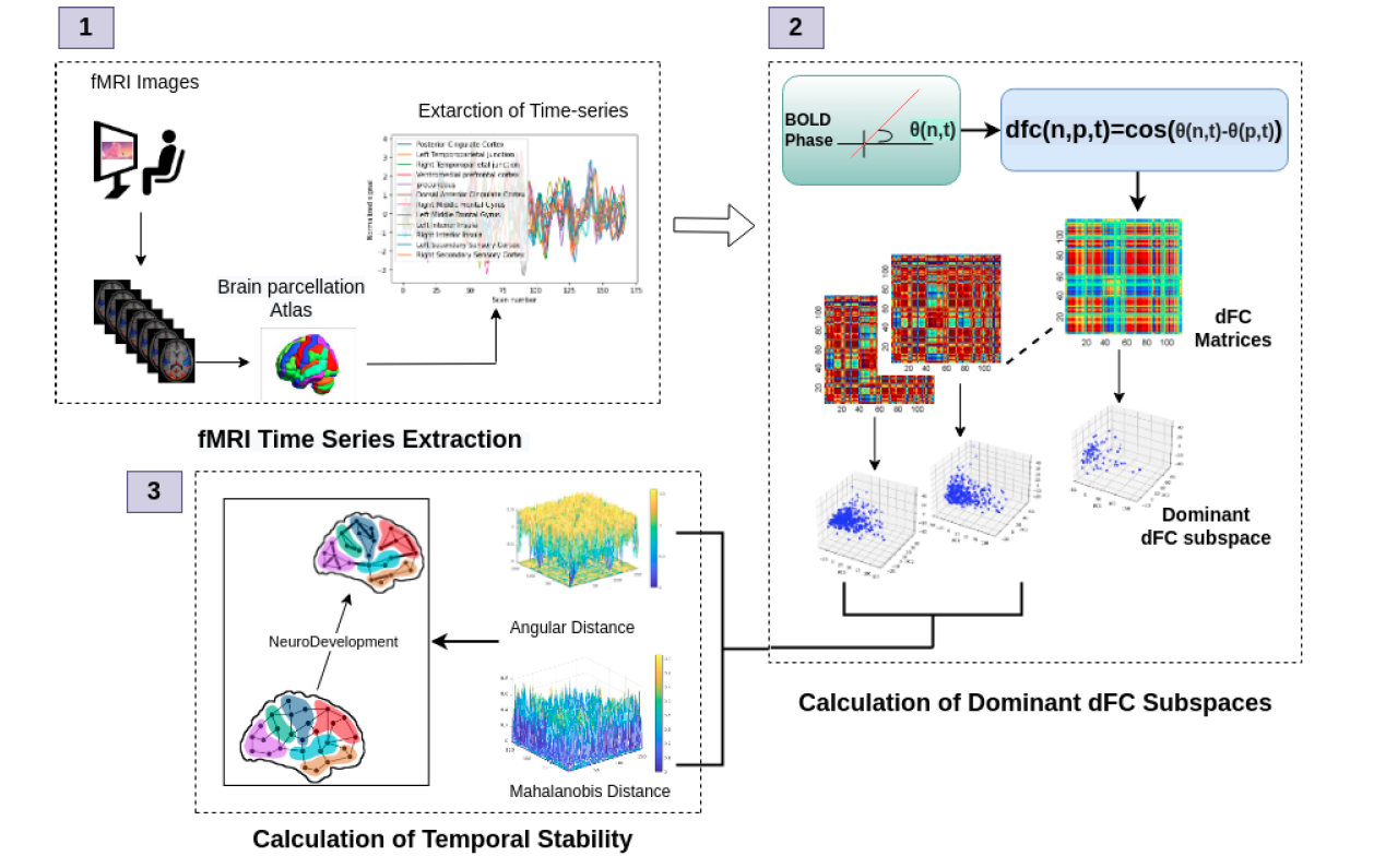
The human brain is a complex system capable of producing non-stationary spatiotemporal signals. Mathematical descriptions based on neural mass models describing population firing rate and time-dependent analysis of regional time series make it possible to predict system dynamics, establishing a direct bridge between biologically inspired theory, simulations, and experimental design. In our lab, we examine structural and functional brain networks using data from non-invasive neuroimaging techniques (fMRI, MEG, MRI, DTI, DSI) to determine fundamental organizational principles of both underlying anatomy and functional dynamics. We are also interested in the emerging field of computational neuropsychiatry, where evidence accumulates from neuropsychiatric disease — specifically schizophrenia and Parkinson’s disease — that exhibits disruption of normal connectivity patterns and neurochemical balance.
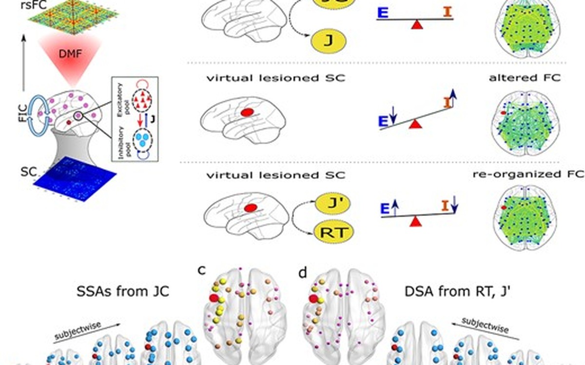
Chakraborty et al.
Cerebral Cortex Communications
2023 · 4(3), tgad012
Structural-and-dynamical similarity predicts compensatory brain areas driving the post-lesion functional recovery mechanism
The focal lesion alters the excitation-inhibition (E-I) balance and healthy functional connectivity patterns, which may recover over time. One possible mechanism for the brain to counter the insult is global reshaping of functional connectivity alterations. However, the operational principles by which this can be achieved remain unknown. We propose a novel equivalence principle based on structural and dynamic similarity analysis to predict whether specific compensatory areas initiate lost E-I regulation after lesion. We hypothesize that similar structural areas (SSAs) and dynamically similar areas (DSAs) corresponding to a lesioned site are the crucial dynamical units to restore lost homeostatic balance within the surviving cortical brain regions. SSAs and DSAs are independent measures, one based on structural similarity properties measured by the Jaccard Index and the other based on post-lesion recovery time. We unravel the relationship between SSA and DSA by simulating a whole-brain mean field model deployed on top of a virtually lesioned structural connectome from human neuroimaging data to characterize global brain dynamics and functional connectivity at the level of individual subjects. Our results suggest that wiring proximity and similarity are the 2 major guiding principles of compensation-related utilization of hemisphere in the post-lesion functional connectivity re-organization process.
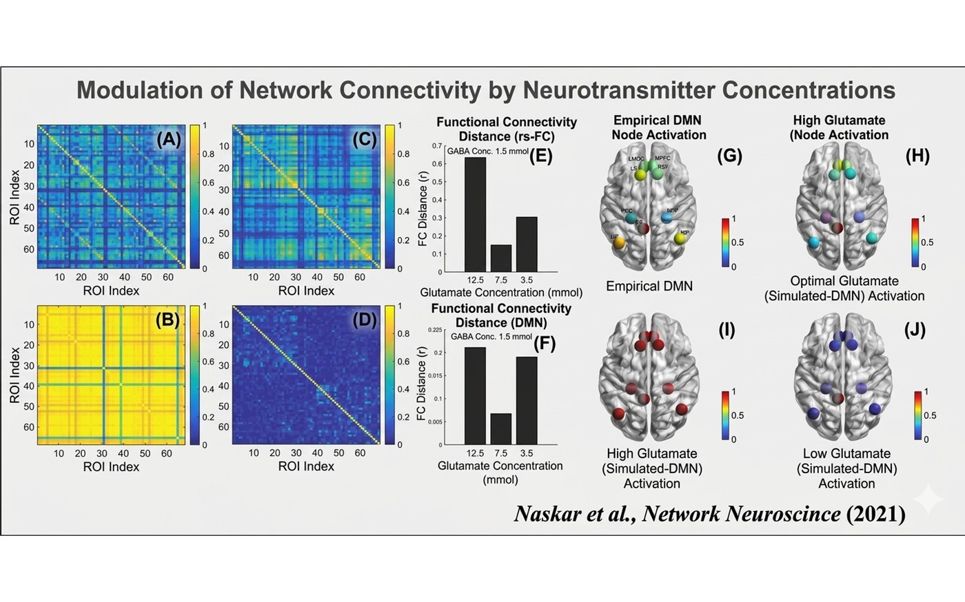

Naskar et al.
Network Neuroscience
2021 · 1-55
Multiscale dynamic mean field model (MDMF) to relate resting-state brain dynamics with local cortical excitatory-inhibitory neurotransmitter homeostasis
Previous computational models have related spontaneous resting-state brain activity with local excitatory–inhibitory balance in neuronal populations. However, how underlying neurotransmitter kinetics associated with E–I balance govern resting-state spontaneous brain dynamics remains unknown. Understanding the mechanisms by virtue of which fluctuations in neurotransmitter concentrations, a hallmark of a variety of clinical conditions, relate to functional brain activity is of critical importance. We propose a multiscale dynamic mean field (MDMF) model—a system of coupled differential equations for capturing the synaptic gating dynamics in excitatory and inhibitory neural populations as a function of neurotransmitter kinetics. Individual brain regions are modeled as a population of MDMF and are connected by realistic connection topologies estimated from diffusion tensor imaging data. First, MDMF successfully predicts resting-state functional connectivity. Second, our results show that the optimal range of glutamate and GABA neurotransmitter concentrations serves as the dynamic working point of the brain, that is, the state of heightened metastability observed in empirical blood-oxygen-level-dependent signals. Third, for predictive validity the network measures of segregation (modularity and clustering coefficient) and integration (global efficiency and characteristic path length) from existing healthy and pathological brain network studies could be captured by simulated functional connectivity from an MDMF model.
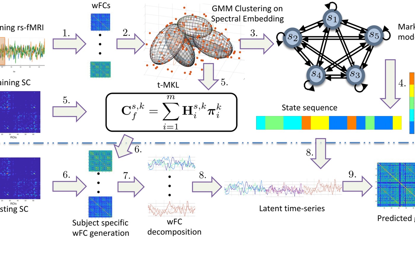

Surampudi et al.
NeuroImage
2019 · 184, 609-620
Resting-state dynamics meets anatomical structure: temporal multiple kernel learning (tMKL) model
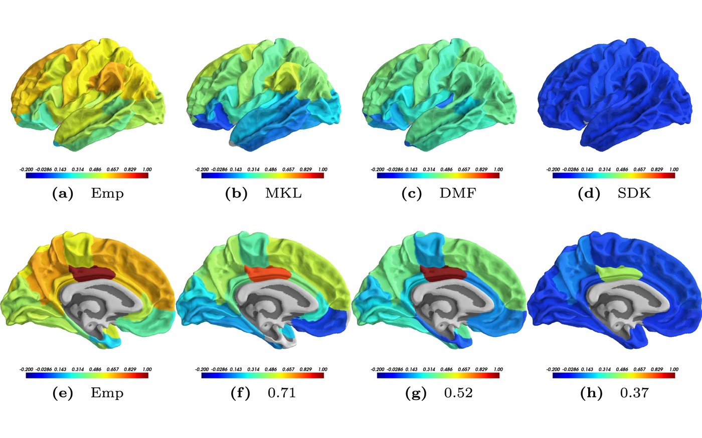
Resting-state functional magnetic resonance imaging (rsfMRI) reveals complex spatio-temporal dynamics in brain activity, and substantial effort has gone into characterizing dynamic functional connectivity (dFC) configurations. Yet the dynamics governing state transitions, and their relationship to stationary functional connectivity (FC), remain an open problem. Existing approaches either characterize dynamics as latent brain states without linking them to structural connectivity (SC), or link dynamic FCs to SC without capturing the temporal evolution of FC. We therefore sought to discover the underlying lower-dimensional manifold representing this temporal structure, parameterized as local density distributions, or latent transient states. We propose temporal Multiple Kernel Learning (tMKL), a graph-theoretic model that learns parameters specific to these states, inherently linking dynamics to structure, and predicts grand average FC via a state transition Markov model. The approach makes no strong assumptions about the data and generalizes to resting or task data for learning subject-specific state transitions, offering a unifying framework for the SC-dFC-FC relationship. Trained and tested on rs-fMRI data from 46 healthy participants, tMKL significantly outperforms existing models — dynamic mean-field (DMF), single diffusion kernel (SDK), and multiple kernel learning (MKL) — at predicting resting-state FC. Validation on an independent cohort of 100 Human Connectome Project participants confirms generalizability. Importantly, tMKL retains sensitivity to subject-specific anatomy, a distinctive contribution toward a holistic approach to SC-FC characterization.
Surampudi et al.
Scientific Reports
2018 · 8(1), 3265
Multiple kernel learning model for relating structural and functional connectivity in the brain
A challenging problem in cognitive neuroscience is to relate the structural connectivity (SC) to the functional connectivity (FC) to better understand how large-scale network dynamics underlying human cognition emerge from the relatively fixed SC architecture. Recent modeling attempts point to the possibility of a single diffusion kernel giving a good estimate of the FC. We highlight the shortcomings of the single-diffusion-kernel model (SDK) and propose a multi-scale diffusion scheme. Our multi-scale model is formulated as a reaction-diffusion system giving rise to spatio-temporal patterns on a fixed topology. We hypothesize the presence of inter-regional co-activations (latent parameters) that combine diffusion kernels at multiple scales to characterize how FC could arise from SC. We formulated a multiple kernel learning (MKL) scheme to estimate the latent parameters from training data. Our model is analytically tractable and complex enough to capture the details of the underlying biological phenomena. The parameters learned by the MKL model lead to highly accurate predictions of subject-specific FCs from test datasets at a rate of 71%, surpassing the performance of the existing linear and non-linear models. We provide an example of how these latent parameters could be used to characterize age-specific reorganization in the brain structure and function.
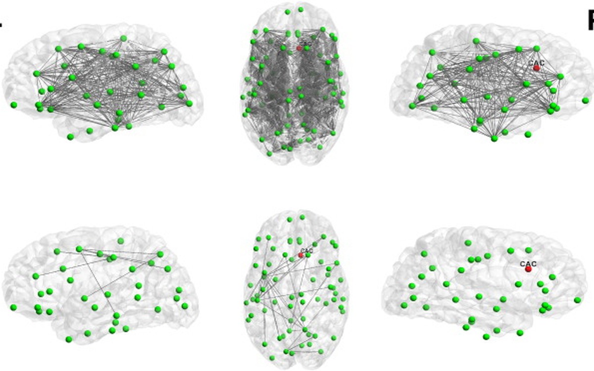

Vattikonda et al.
NeuroImage
2016 · 136, 57-67
Does the regulation of local excitation-inhibition balance aid in recovery of functional connectivity? A computational account
Computational modeling of the spontaneous dynamics over the whole brain provides critical insight into the spatiotemporal organization of brain dynamics at multiple resolutions and their alteration to changes in brain structure (e.g., in diseased states, aging, across individuals). Recent experimental evidence further suggests that the adverse effect of lesions is visible on spontaneous dynamics characterized by changes in resting state functional connectivity and its graph theoretical properties (e.g., modularity). These changes originate from altered neural dynamics in individual brain areas that are otherwise poised towards a homeostatic equilibrium to maintain a stable excitatory and inhibitory activity. In this work, we employ a homeostatic inhibitory mechanism, balancing excitation and inhibition in the local brain areas of the entire cortex under neurological impairments like lesions, to understand global functional recovery (across brain networks and individuals). Previous computational and empirical studies have demonstrated that the resting state functional connectivity varies primarily due to the location and specific topological characteristics of the lesion. We show that local homeostatic balance provides a functional recovery by re-establishing excitation–inhibition balance in all areas that are affected by the lesion. We systematically compare the extent of recovery in the primary hub areas (e.g., default mode network (DMN), medial temporal lobe, medial prefrontal cortex) as well as other sensory areas like primary motor area, supplementary motor area, fronto-parietal and temporo-parietal networks. Our findings suggest that stability and richness similar to the normal brain dynamics at rest are achievable by re-establishment of balance.
Sensory Processing, Perception, Attention, Predictive Coding, and Learning in Memory and Cognition
Behaviour, EEG, MEG, fMRI, and deep neural networks
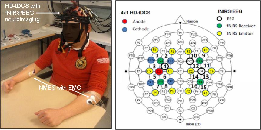
We aim to investigate the role of local oscillations in a normal human brain, and the functional role of abnormal oscillations in neuropsychiatric disorders characterised by alterations in distributed activity across brain areas. We use network methods to uncover changes in large-scale brain circuitry that impact cognitive function and behaviour, to identify the underlying neurophysiological processes of disease, and inform clinical interventions. A related focus is critical behaviour in resting-state dynamics: the precise neuronal mechanism generating near-critical dynamics in the brain remains unresolved despite numerous recent investigations. To mathematically describe empirical observations, we study cortical long-range correlations in space and time, and for short-range correlations, we examine features such as neuronal avalanches. We also develop drift-diffusion models based on statistical physics to study the mesoscopic dynamics of neural masses distributed across graph-like entities such as voxels and nodes connected via realistic structural connectivity matrices.
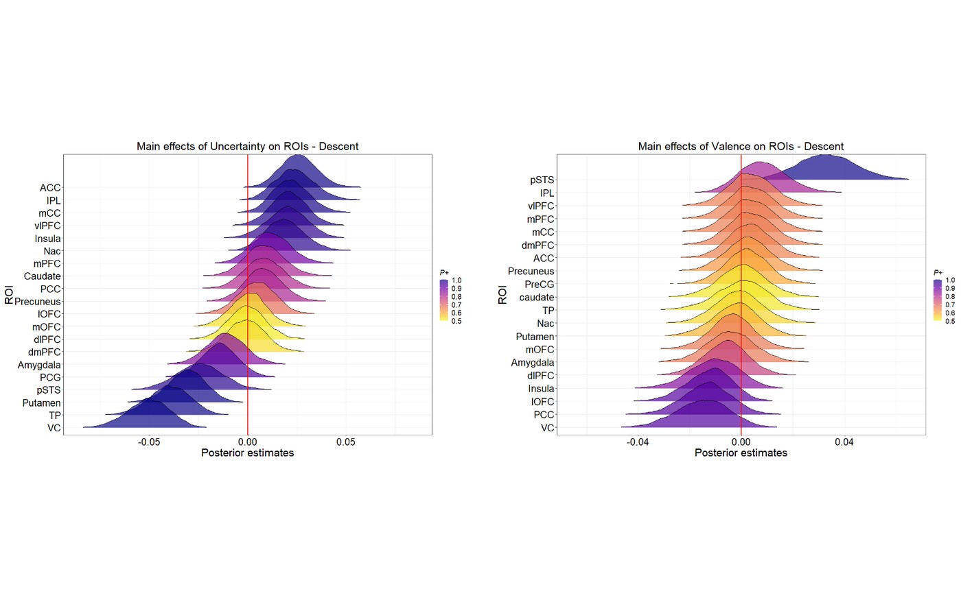
Majumdar et al.
Cerebral Cortex
2023 · 33(7), 3750-3772
Emotion dynamics as hierarchical Bayesian inference in time
What fundamental property of our environment would be most valuable and optimal in characterizing the emotional dynamics we experience in daily life? Empirical work has shown that an accurate estimation of uncertainty is necessary for our optimal perception, learning, and decision-making. However, the role of this uncertainty in governing our affective dynamics remains unexplored. Using Bayesian encoding, decoding and computational modeling on a large-scale neuroimaging and behavioral data on a passive movie-watching task, we showed that emotions naturally arise due to ongoing uncertainty estimations about future outcomes in a hierarchical neural architecture. Several prefrontal subregions hierarchically encoded a lower-dimensional signal that highly correlated with the evolving uncertainty. Crucially, the lateral orbitofrontal cortex (lOFC) tracked the temporal fluctuations of this uncertainty and was predictive of the participants' predisposition to anxiety. Furthermore, we observed a distinct functional double-dissociation within OFC with increased connectivity between medial OFC and DMN, while with that of lOFC and FPN in response to the evolving affect. Finally, we uncovered a temporally predictive code updating an individual's beliefs spontaneously with fluctuating outcome uncertainty in the lOFC. A biologically relevant and computationally crucial parameter in the theories of brain function, we propose uncertainty to be central to the definition of complex emotions.

Kumar et al.
Neuropsychologia
2023 · 184, 108559
Effective networks mediate right hemispheric dominance of human 40 Hz auditory steady-state response
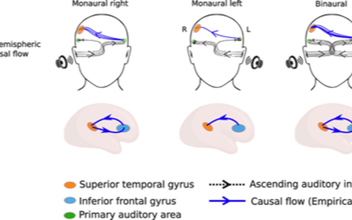
Auditory steady-state responses (ASSR) are induced from the brainstem to the neocortex when humans hear periodic amplitude-modulated tonal signals. ASSRs have been argued to be a key marker of auditory temporal processing and pathological reorganization of ASSR, a biomarker of neurodegenerative disorders. However, most of the earlier studies reporting the neural basis of ASSRs were focused on looking at individual brain regions. Here, we seek to characterize the large-scale directed information flow among cortical sources of ASSR entrained by 40 Hz external signals. Entrained brain rhythms with power peaking at 40 Hz were generated using both monaural and binaural tonal stimulation. First, we confirm the presence of ASSRs and their well-known right hemispheric dominance during binaural and both monaural conditions. Thereafter, reconstruction of source activity employing individual anatomy of the participant and subsequent network analysis revealed that while the sources are common among different stimulation conditions, differential levels of source activation and differential patterns of directed information flow among sources underlie processing of binaurally and monaurally presented tones. Particularly, we show that bidirectional interactions involving the right superior temporal gyrus and inferior frontal gyrus underlie right hemispheric dominance of 40 Hz ASSR during both monaural and binaural conditions. On the other hand, for monaural conditions, the strength of inter-hemispheric flow from left primary auditory areas to right superior temporal areas followed a pattern that complies with the generally observed contralateral dominance of sensory signal processing.
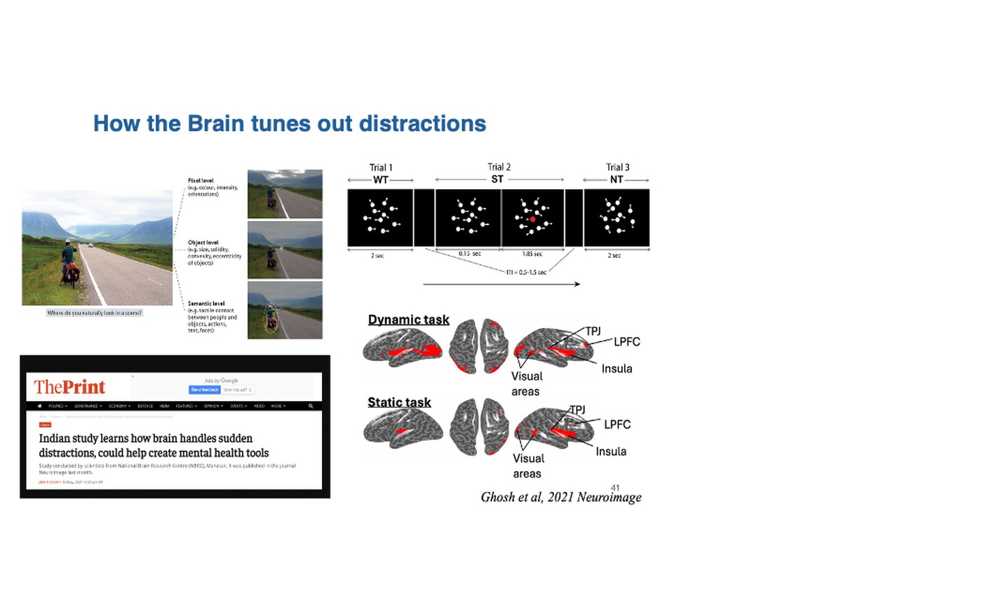

Ghosh et al.
NeuroImage
2021 · 231, 117869
Organization of directed functional connectivity among nodes of ventral attention network reveals the common network mechanisms underlying saliency processing across distinct spatial and spatio-temporal scales
Previous neuroimaging studies have extensively evaluated the structural and functional connectivity of the Ventral Attention Network (VAN) and its role in reorienting attention in the presence of a salient (pop-out) stimulus. However, a detailed understanding of the "directed" functional connectivity within the VAN during the process of reorientation remains elusive. Functional magnetic resonance imaging (fMRI) studies have not adequately addressed this issue due to a lack of the appropriate temporal resolution required to capture this dynamic process. The present study investigates the neural changes associated with processing salient distractors operating at a slow and a fast time scale using a custom-designed experiment involving visual search on static images and dynamic motion tracking, respectively. We recorded high-density scalp electroencephalography (EEG) from healthy human volunteers, obtained saliency-specific behavioral and spectral changes during the tasks, localized the sources underlying the spectral power modulations with individual-specific structural MRI scans, reconstructed the waveforms of the sources, and finally, investigated the causal relationships between the sources using spectral Granger-Geweke Causality (GGC). We found that salient stimuli processing, across tasks with varying spatio-temporal complexities, involves a characteristic modulation in the alpha frequency band which is executed primarily by the nodes of the VAN constituting the temporo-parietal junction (TPJ), the insula, and the lateral prefrontal cortex (lPFC). The directed functional connectivity results further revealed the presence of bidirectional interactions among prominent nodes of right-lateralized VAN, corresponding only to the trials with saliency. Thus, our study elucidates the invariant network mechanisms for processing saliency in visual attention tasks across diverse time-scales.
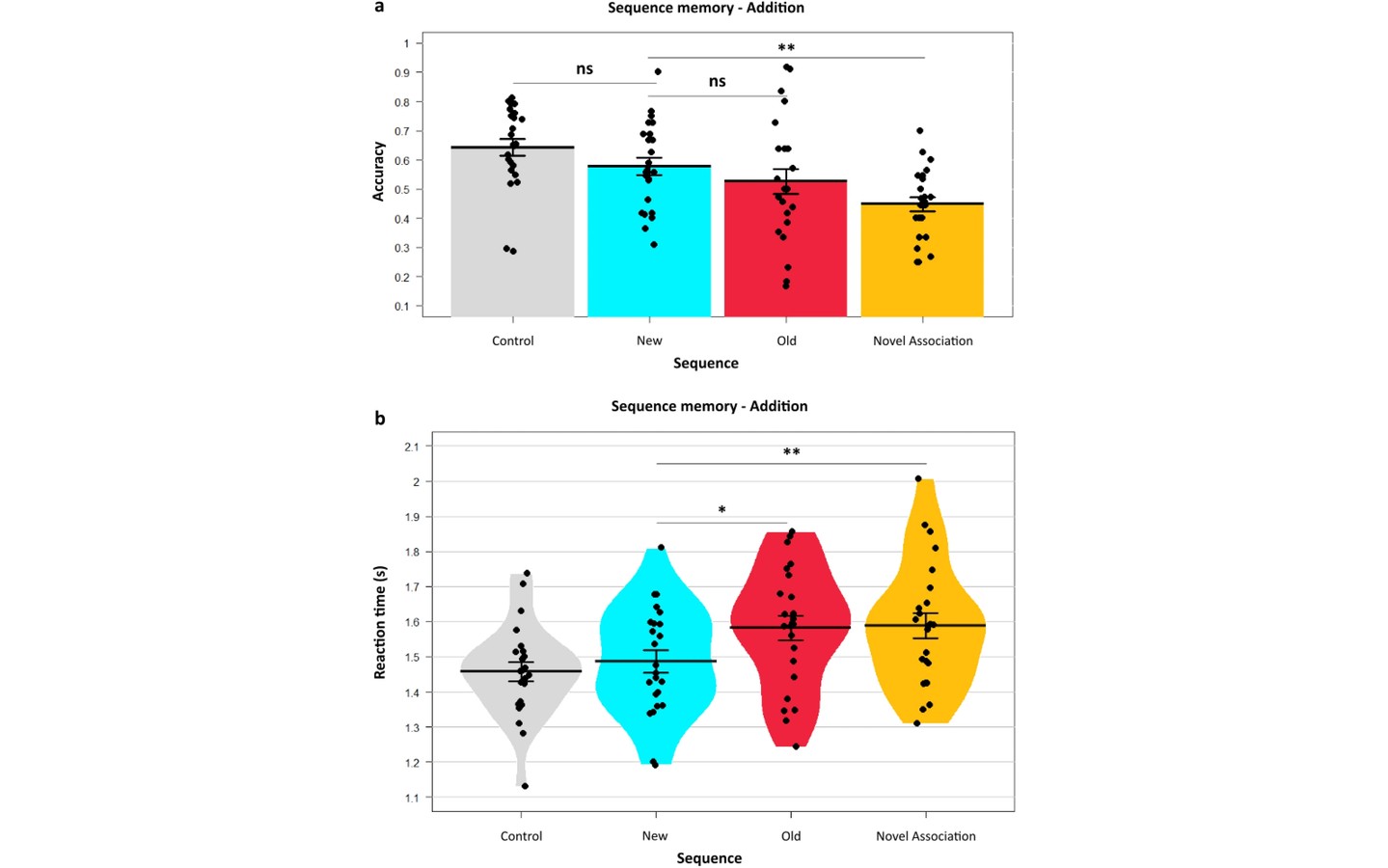

Yazin et al.
Scientific Reports
2021 · 11(1), 12364
Contextual prediction errors reorganize naturalistic episodic memories in time
Episodic memories are contextual experiences ordered in time. This is underpinned by associative binding between events within the same contexts. The role of prediction errors in declarative memory is well established, but has not been investigated in the time dimension of complex episodic memories. Here, we combine these two properties of episodic memory, extend them into the temporal domain, and demonstrate that prediction errors in different naturalistic contexts lead to changes in the temporal ordering of event structures in them. The wrongly predicted older sequences were weakened despite their reactivation. Interestingly, the newly encoded sequences with prediction errors, seen once, showed accuracy as high as control sequences, which were viewed repeatedly without change. Drift–diffusion modelling revealed a lower decision threshold for the newer sequences than the older sequences, reflected by their faster recall. Moreover, participants' adjustments to their decision threshold significantly correlated with their relative speed of sequence memory recall. These results suggest a temporally distinct and adaptive role for prediction errors in learning and reorganizing episodic temporal sequences.
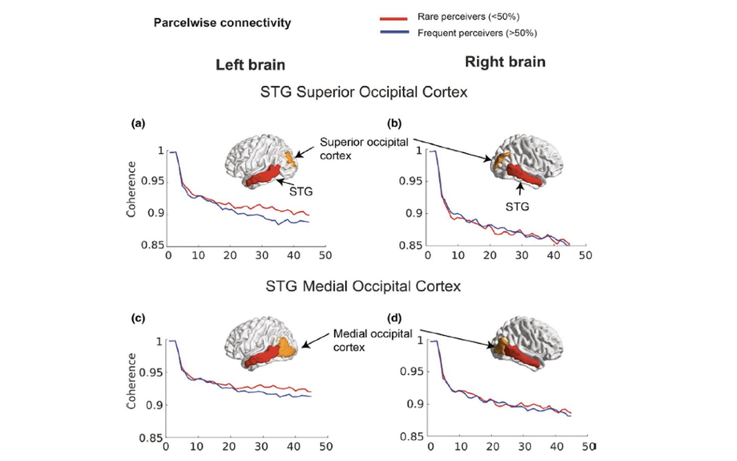

Kumar et al.
European Journal of Neuroscience
2020 · 52(7), 3746-3762
Biophysical mechanisms governing large-scale brain network dynamics underlying individual-specific variability of perception
Perception necessitates interaction among neuronal ensembles, the dynamics of which can be conceptualized as the emergent behavior of coupled dynamical systems. Here, we propose a detailed neurobiologically realistic model that captures the neural mechanisms of inter-individual variability observed in cross-modal speech perception. From raw EEG signals recorded from human participants when they were presented with speech vocalizations of McGurk-incongruent and congruent audio-visual (AV) stimuli, we computed the global coherence metric to capture the neural variability of large-scale networks. We identified that participants' McGurk susceptibility was negatively correlated with their alpha band global coherence. The proposed biophysical model conceptualized the global coherence dynamics that emerge from coupling between the interacting neural masses, representing the sensory-specific auditory/visual areas and modality nonspecific associative/integrative regions. Subsequently, we could predict that an extremely weak direct AV coupling results in a decrease in alpha band global coherence, mimicking the cortical dynamics of participants with higher McGurk susceptibility. Source connectivity analysis also showed decreased connectivity between sensory-specific regions in participants more susceptible to the McGurk effect, thus establishing an empirical validation of the prediction. Overall, our study provides an outline to link variability in structural and functional connectivity metrics to variability of performance that can be useful for several perception and action task paradigms.
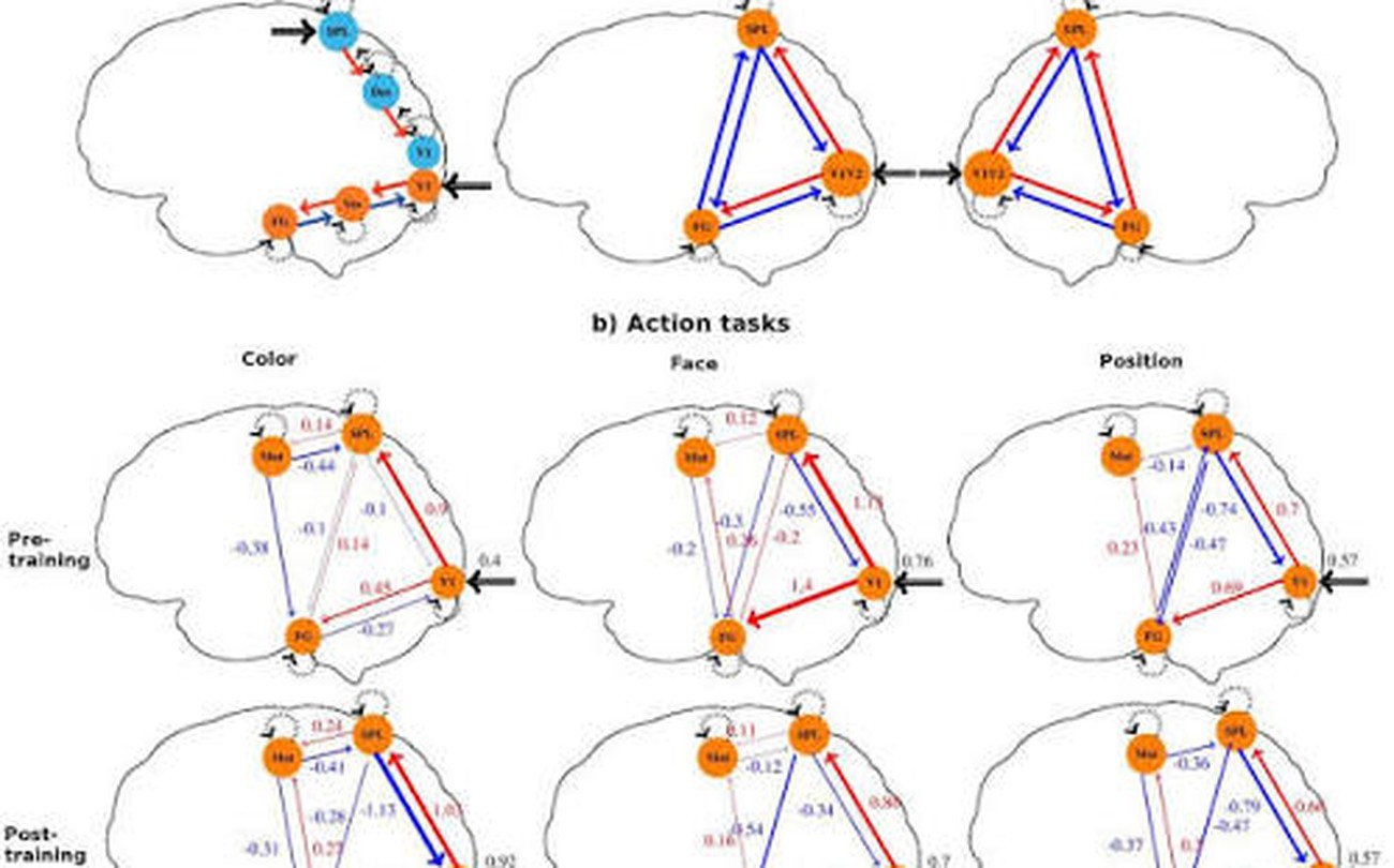
Ray et al.
Journal of Cognitive Neuroscience
2020 · 1-15
Large-scale functional integration, rather than functional dissociation along dorsal and ventral streams, underlies visual perception and action
Visual dual-stream theory posits that two distinct neural pathways of specific functional significance originate from primary visual areas and reach the inferior temporal (ventral) and posterior parietal areas (dorsal). However, there are several unresolved questions concerning the fundamental aspects of this theory. For example, is the functional dissociation between ventral and dorsal stream driven by features in input stimuli, or is it driven by categorical differences between visuoperceptual and visuomotor functions? Is the dual stream rigid or flexible? What is the nature of the interactions between the two streams? We addressed these questions using fMRI recordings on healthy human volunteers and employing stimuli and tasks that can tease out the divergence between visuoperceptual and visuomotor variants of dual-stream theory. fMRI scans were repeated after seven practice sessions that were conducted in a non-MRI environment to investigate the effects of neuroplasticity. Brain activation analysis supports an input-based functional dissociation and the existence of context-dependent neuroplasticity in dual-stream areas. Intriguingly, premotor cortex activation was observed in the position perception task, and distributed deactivated regions were observed in all perception tasks, thus warranting a network-level analysis. Dynamic causal modeling analysis incorporating activated and deactivated brain areas during perception tasks indicates that the brain dynamics during visual perception and actions could be interpreted within the framework of predictive coding. Effectively, the network-level findings point toward the existence of more intricate context-driven functional networks selective of "what" and "where" information rather than segregated streams of processing along ventral and dorsal brain regions.
Funding
- Science and Engineering Research Board (SERB) Core Research Grant, Govt. of India
- Department of Biotechnology (DBT) Flagship project on Common Mental Health and Brain Mapping
- Department of Biotechnology (DBT), Govt. of India
- Department of Science and Technology (DST) Cognitive Science Research Initiative, Govt. of India
- Department of Science and Technology (DST)
- NBRC and IIT Jodhpur core